Digestive System Basics
|
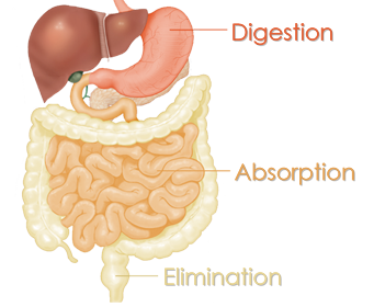
DIGESTIONChemical and mechanical breakdown of foods into smaller units that can be taken across the intestinal epithelium into the body. |
ABSORPTIONActive or passive transfer of digested nutrients from the lumen of the GI tract to the extracellular fluid. Circulation brings nutrients to every cell in the body, which are used by the cells as energy to do work. |
ELIMINATIONUndigested waste material leaves the body. |
|
3 chief functions
|
The digestive system can be broken down into two groups of organs:
Digestive Tract
- Digestive tract (also referred to as gastrointestinal or GI tract and rarely, the alimentary tract)
- Continuous passageway beginning at the mouth, where food is taken in, and terminating at the anus, where solid waste products of digestion leave the body
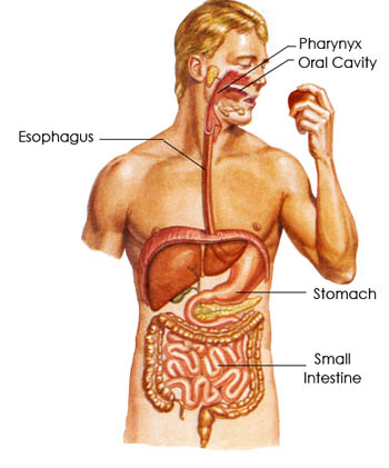
- Muscular tube extending throughout body that includes: mouth, pharynx, esophagus, stomach, small intestine, and large intestine
The Oral Cavity
The oral cavity , or mouth, is the starting point of a substance as it travels through the GI tract. It digests food both mechanically and chemically.
- Mechanical:
- Ingestion – receives food
- Mastication (chewing) – breaks food into smaller pieces with teeth
- Deglutition – moves proper amount of food toward throat to be swallowed
- Chemical:
- Saliva is produced by salivary glands and released into the oral cavity, lubricates food and begins starch digestion with digestive enzyme, salivary amylase (more specifically ptyalin, a type of alpha-amylase)
- Salivary glands also secrete lysozyme, which kills bacteria attempting to enter the digestive system
The Pharynx
The pharynx (commonly referred to as the throat)
- Anatomy:
- Oropharynx – oral part, visible when you open mouth and depress tongue
- Palatine tonsils are on either side of the oropharynx
- Nasopharynx – section extending up toward nasal cavity
- Laryngeal pharynx – section extending down to larynx
- Soft plate forms posterior of oral cavity
- Uvula – conical projection of connective tissue that hangs from soft plate
- During eating the tongue pushes a small bolus of food and saliva into pharynx and swallowing occurs by involuntary reflex action
- The soft plate and uvula raise to prevent food/liquid from entering the nasopharynx
- The entrance of the trachea is similarly guarded by the epiglottis, a leaf-shaped cartilage
The Esophagus
The esophagus is a muscular tube, about 10 inches long.
- No digestion occurs in this section of the GI tract
- Food is lubricated with mucus and moved by peristalsis into the stomach
- Peristalsis = alternating muscle contractions in the esophageal walls create wavelike movements to propel food through the GI tract and mix it with digestive juices
- Passes through diaphragm before joining the stomach
The Stomach
The stomach is a bag-like organ that can hold up to 2 liters of food and fluid when fully expanded.
- Anatomy:
- J-shaped, located in upper left region of abdominal cavity
- Extra layer of smooth muscle in wall aids in grinding and mixing food
- Rugae – inner lining of stomach forms numerous folds when empty
- Lower esophageal sphincter (LES) separates esophagus and stomach, also known as cardiac sphincter
- Pyloric sphincter separates stomach and small intestine
- Pylorus = region of stomach leading into sphincter, controls how rapidly food travels to small intestine
- Cells lining stomach secrete substances that mix together to form gastric juice
- Gastric juice:
- Hydrochloric acid (HCl) = strong acid that helps break down proteins and destroy foreign organisms
- Pepsin = enzyme that digests proteins, produced in inactive form and activated only when food enters stomach and HCl is present
- Some cells also secrete a substantial amount of mucus to protect stomach lining from digestion secretions
- Also secretes fat-digesting enzyme, lipase, but not important in adults
- Chyme = highly acidic, semi-liquid mixture of gastric juice and food that leaves stomach to enter small intestine
The Small Intestine
The small intestine is the longest part of digestive tract but smaller in diameter than large intestine
- Anatomy:
- Approximately 3 meters long (10 feet) in life (can be up to 6 meters stretched out after death) with an average width of 2.5 centimeters (1 inch)
- The organ is divided into three sections.
- The duodenum is the first 25 centimeters (10 inches)
- Two innermost layers of digestive tract (called the mucosa and submucosa) contain glands that excrete large amounts of mucus to protect the small intestine from strongly acidic chyme entering from stomach
- Digestive juices from the liver and pancreas enter through a small opening
- Most digestion occurs here under the effects of these juices
- The jejunum is the next two fifths of the intestine
- The ileum is the remaining portion
- Mucosal cells secrete enzymes that digest proteins and carbohydrates
- Produce maltase, sucrase, and lactase
- Act on the disaccharides of maltose, sucrose, and lactose respectively to convert them to simpler, more absorbable forms
- Peptidases – digests proteins to amino acids
- Most absorption of digested food, water, and minerals occurs through the walls of the small intestine
- Mucosa, inner layer of lumen, forms fingerlike projections called villi, which in turn contain cells with projecting folds of plasma membrane called microvilli
- Increases surface area of small intestine dramatically, aiding in effective absorption of digested nutrients across membrane wall
- Each villus contains blood vessels through which most digestion products are absorbed into the blood and a lacteal (specialized lymphatic capillary) through which fats are absorbed into the lymph
Accessory Organs
- Accessory organs – necessary for digestion but not a direct part of the digestive tract, rather they secrete substances into the digestive tract through ducts
- Includes: salivary glands, liver, gallbladder, and pancreas
The Large Intestine
The large intestine is approximately 1.5 meters long (5 feet) and 6.5 centimeters (2.5 inches) in diameter
- Anatomy:
- The cecum is the first part
- The ileocecal valve divides the ileum of the small intestine and the cecum
- Prevents food from traveling backward into small intestine
- Vermiform appendix (aka the appendix) is a small blind tube containing lymphoid tissue and attached to the cecum
- In order, the subdivisions are:
- Ascending colon, transverse colon, descending colon, sigmoid colon, rectum, and anal canal
- Rectum = temporary storage area for indigestible or non-absorbable food residue
- Anal canal leads to outside of body through anus
- Large intestine secretes mucus but no digestive enzymes
- Food is not digested
- Water is reabsorbed, undigested food is stored, formed into solid waste material (feces) and eliminated
- During food residue storage, symbiotic (“good”) bacteria act on it to produce vitamin K and some of the B-complex vitamins
- Antibiotics have the ability to destroy these symbiotic bacteria
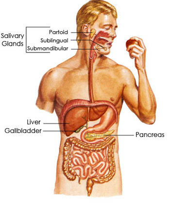
The Salivary Glands
The salivary glands secrete saliva:
- In the mouth, food is mixed with saliva, which moistens the food and facilitates mastication (chewing) and deglutition (swallowing). Helps to keep teeth and mouth clean and contains:
- Some antibodies
- Lysozyme – enzyme that helps reduce bacterial growth
- Mucus
- Salivary amylase – begins digestive process by converting starch to sugar
- Saliva is manufactured by three sets of glands: the parotid glands, submandibular or submaxillary glands, and sublingual glands (located inferior and anterior to the ear, near body of lower jaw, and under tongue respectively)
- All three empty into oral cavity
The Liver
The liver is the body’s largest glandular organ:
- Manufactures bile, a substance needed for digestion of fats
- Contains salts that emulsify fat (break down into small droplets that can be more easily acted on by digestive enzymes)
- Aids in fat absorption in the small intestine
- Leaves liver via two ducts that merge to form the common hepatic duct, collects bile from the gallbladder and the common bile duct delivers bile to the duodenum
- Storage of glucose in the form of glycogen
- When blood sugar levels falls, liver cells convert glycogen into glucose, which is released into the blood
- Modification of fats so that they can be used more efficiently by the body
- Storage of some vitamins and iron
- Formation of blood plasma proteins, like albumin, globulins, and clotting factors
- Destruction of old red blood cells
- Synthesis of urea
- Detoxification of harmful substances like alcohol and drugs
The Gallbladder
The gallbladder is a muscular sac on the inferior surface of the liver that stores bile
- Manufactures bile continuously, but only releases it into the common bile duct when chyme enters the duodenum
The Pancreas
The pancreas is a long gland that extends from the duodenum to the spleen
- Produces enzymes that digest fats, proteins, carbohydrates, and nucleic acids
- Protein-digesting enzymes produced in inactive forms and converted to active forms in the small intestine
- Releases a substantial amount of sodium bicarbonate to neutralize the acidic chyme in the small intestine
- Juices collect in main duct that empty into the common bile duct or into the duodenum near the common bile duct
- Pancreatic juice contains:
- Lipase – digests almost all of the fat that the bile has previously emulsified
- Fats broken down into glycerol and fatty acids, which are more absorbable
- If pancreatic lipase is absent, fats are expelled with feces in undigested form
- Amylase – converts starch into sugar
- Trypsin – splits proteins into smaller units (amino acids), which are small enough to be absorbed through walls of intestine
- Nucleases – enzymes that digest nucleic acids DNA and RNA
- When pancreatic juice absent, serious digestive disturbances always occur
- Also functions as an endocrine gland
- Produces the hormones, insulin and glucagon, which are released into the blood to regulate sugar metabolism
|
|


 For over 14 years, we have made it our mission to collaborate with innovative practitioners, nutritional experts, and scientists to realize their concepts and ideas into life changing products and applications. This cultivated group called "The Founding Scientists," is what makes GHT the unique, robust and respected organization in industries of nutrition and health.